Anaplasmosis
Overview
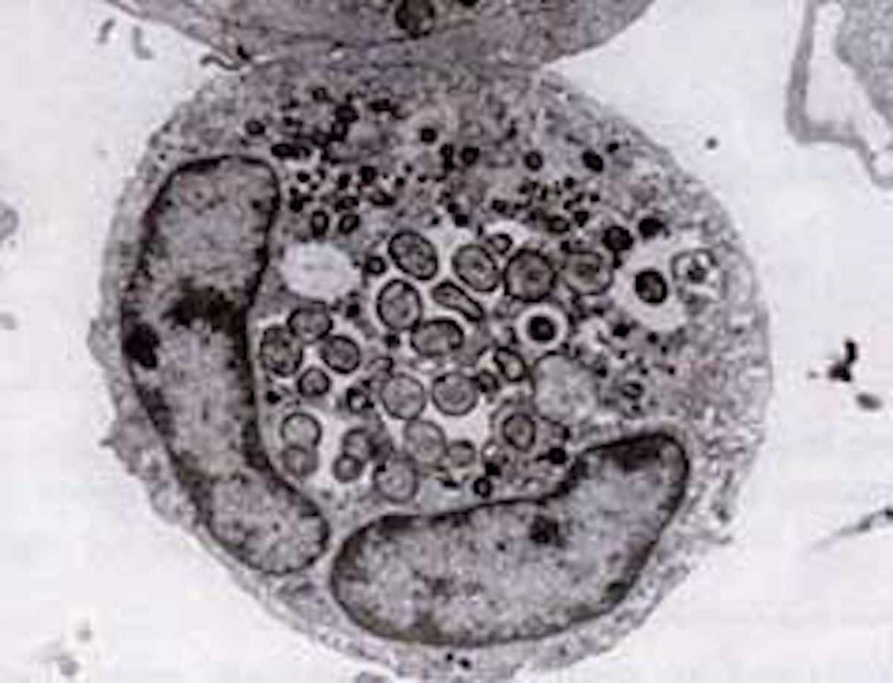
The first case of human anaplasmosis was described in 1990, when a Wisconsin patient developed a severe febrile illness following a tick bite and died two weeks later. Blood smears revealed clusters of bacteria within the patient’s neutrophils, similar to the morulae seen in monocytes with E. chaffeensis infection. However, cultures and serologic tests for E. chaffeensis were negative. Nevertheless, the patient’s clinical course suggested ehrlichiosis of some kind, and when several additional cases of the disease were reported in the northern Midwest in ensuing years, it was posited that a new species of Ehrlichia might be emerging. The new disease was tentatively given the name “human granulocytic ehrlichiosis,” or HGE.
In 1994, DNA sequencing studies revealed that the HGE agent was clearly distinct from E. chaffeensis but essentially identical to two previously known ehrlichial veterinary pathogens, E. equi and E. (Cytoecetes) phagocytophila. Under a new taxonomic scheme since implemented, these three organisms have been united as a single species within a new genus, Anaplasma. The new species is referred to as Anaplasma phagocytophilum, and the disease it causes is now known as human granulocytic anaplasmosis, or HGA.
Like Ehrlichia species, Anaplasma organisms are small, gram-negative, and intracellular. A. phagocytophilum targets neutrophils, alters their function in the host, and forms morulae within vacuoles. Most of the damage it causes appears to be related to host inflammatory processes, as there is little evidence of a correlation between the number of organisms and host disease severity.
Anaplasmosis is a global infection, occurring in North America, most of Europe, and eastern Asia. Ticks from the Ixodes persulcatus-complex are the vectors: I. scapularis in the northeastern and upper Midwestern regions of the United States; I. pacificus in the Pacific Northwest; I. ricinus in Europe; and I. persulcatus in Asia. A. phagocytophilum is maintained in nature by cycling between these ticks and various small mammals, primarily mice and other small rodents. Because Ixodes ticks are also the vectors for Lyme disease, babesiosis, and tick-borne encephalitis, Anaplasma coinfection with these other diseases can and does occur in humans.
In the United States, cases of HGA have outnumbered those of Human Monocytic Ehrlichiosis (HME) Similar to HME and human ewingii ehrlichiosis, the median age of patients with HGA is around 50 years old.
Signs and Symptoms
The clinical course of HGA ranges from asymptomatic infection to fatal disease. Initial symptoms, occuring about five to ten days after tick bite, are largely non-specific: fever, chills, severe headache, and myalgias;j nausea, cough, and arthralgias may also occur. Rash is uncommon but has been reported.
Compared with HME, HGA appears less likely to involve the central nervous system, but peripheral neuropathies (e.g., numbness and tingling) are more common and can last weeks to months. Among the neurologic findings reported in the medical literature are facial palsy, demyelinating polyneuropathy, and brachial plexopathy. Respiratory distress syndrome and a septic or toxic shock-like syndrome have been reported, but appear to be less common than in HME. The overall fatality rate from HGA is very low (less than 1%), with most of the deaths resulting from opportunistic infections (for example, herpes simplex esophagitis, Candida pneumonitis, and pulmonary aspergillosis) in immunocompromised patients.
Diagnosis
Standard blood tests in HGA usually reveal findings similar to those seen in HME: leukopenia, thrombocytopenia, and liver function abnormalities (elevated transaminases). However, the hematological abnormalities frequently resolve by the second week of symptoms, so their absence should be interpreted in that context if patients are presenting later in the course of their illness. In general, empiric antibiotic treatment should be considered for patients in endemic areas who present with an acute febrile illness suggestive of HGA.
For specific diagnosis, Wright or Giemsa-stained blood smears have a slightly higher yield than with HME, but are still not optimal for general clinical utility, given that there appears to be a wide variation (25-75%) in the sensitivity of these tests in visualizing morulae in host neutrophils. More helpful, but not always available, are polymerase chain reaction (PCR) tests, which are estimated to have a sensitivity of 67-90%. Prior antibiotic therapy dramatically reduces the sensitivity of both of these diagnostic methods.
Serologic testing is useful to confirm the diagnosis of anaplasmosis. The most commonly used method is indirect immunofluorescence (IFA) of IgM and IgG anti-A. phagocytophilum antibodies. Seroconversion is perhaps the most sensitive laboratory evidence of A. phagocytophilum infection, but is not always obtained in a timely enough manner to provide useful input on clinical (i.e., treatment) decisions.